Introduction:
“stroke” is a in medical terminology refers to a sudden interruption of blood supply to the brain, resulting in the rapid loss of brain function.
This interruption can be caused by a blockage of blood flow (ischemic stroke) or the rupture of a blood vessel (hemorrhagic stroke). As a result, the affected part of the brain may not receive enough oxygen and nutrients, leading to the death of brain cells.
Etiology:
When discussing the etiology of it, there are several factors and conditions that can contribute to its occurrence.
Emboli
Hemorrhage
Ischemia
Hypertension
Atherosclerosis
Risk factor:
High Blood Pressure: This is the leading risk factor for stroke. Consistently high blood pressure can damage blood vessels in the brain, making them more likely to rupture or become blocked (>160/90mm Hg)
Smoking: Smoking contributes to the buildup of plaque in the arteries, increasing the risk of stroke.
Diabetes: Diabetes can increase the risk of stroke by contributing to the buildup of fatty deposits in blood vessels.
High Cholesterol: High levels of low-density lipoprotein (LDL) cholesterol can lead to the formation of plaques in the arteries, which can restrict blood flow to the brain.
Heart Disease: Conditions like atrial fibrillation, heart valve disease, and other heart problems can increase the risk of stroke.
Obesity: Being overweight or obese can increase the risk of high blood pressure, diabetes, and high cholesterol, all of which contribute to stroke risk.
Physical Inactivity: Lack of exercise can contribute to obesity, high blood pressure, and other health issues that increase stroke risk.
Excessive Alcohol Consumption: Drinking large amounts of alcohol can increase blood pressure and contribute to other health issues that raise stroke risk.
Warning signs:
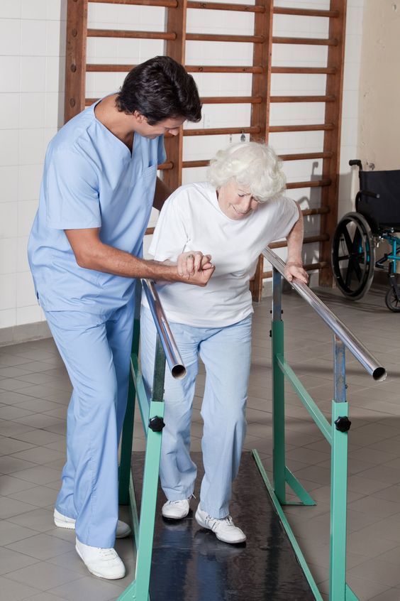
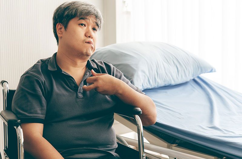
PHYSIOTHERAPY MANAGEMENT:
Constraint-Induced Movement Therapy (CIMT): Involves constraining the unaffected limb to encourage use of the affected limb.
Bilateral Training: Exercises that involve both sides of the body to improve motor control and coordination.
Simulated Activities: Practicing real-life tasks in a controlled environment to improve functional outcomes.
Cognitive and Perceptual Training: Addressing any cognitive or perceptual deficits that impact motor function.
Patient and Family Education: Providing information about stroke recovery, home exercises, and ways to manage stroke-related challenges.
Support and Motivation: Encouraging patients and their families throughout the rehabilitation process.
conclusion:
It is a medical emergency that demands immediate attention. Early recognition and timely treatment can significantly reduce brain damage and disability. With proper rehabilitation, lifestyle changes, and continuous care, many individuals can regain independence and improve quality of life.